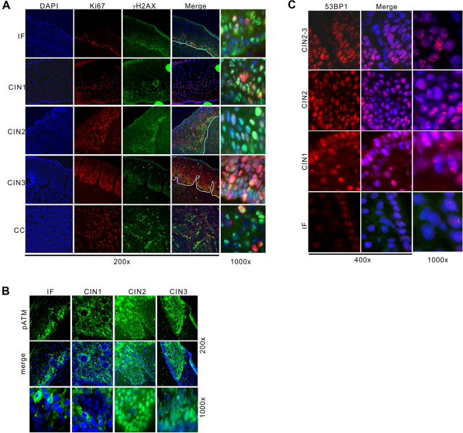
Fig. 1.
Elevated DNA damage in cervical epithelium. A Indirect immunofluorescent staining for Ki67, γH2AX in cervical epithelium. Nuclei were counterstained by DAPI. Stages of CIN and cervical cancer and magnifications (200 × and 1000x) are indicated. IF: inflammatory lesion. B, C As in (A), immunostaining for phosphorylated ATM (pS1981, B) and 53BP1 (C) in nuclear compartment of cervical epithelial cells. pS1981 displayed cytoplasmic staining in inflammatory lesions and CIN I, which we regarded as non-specific. All cervical samples were diagnosed as HPV16-positive according to pathological examination (see methods)

