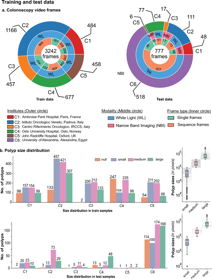
Figure 1.
Multi-centre training and test samples. (a) Colonoscopy video frames for which the annotation samples were reviewed and released as training (left) and test (right) are provided. Training samples included nearly proportional frames from five centres (C1–C5). In contrast, test samples consisted of a majority of single and sequence frames from the unseen centre (C6) with white light modality (WL) only. Test data from the seen centres C1, C3, and C5 consisted of only NBI images, while centres C2 and C4 consisted of white light (WL) and narrow-band imaging (NBI) modalities. (b) The number of polyp counts and samples with no polyps per centre are provided. Polyp sizes (in pixels) were classified based on resized image frames of pixels. Polyp sizes (in pixels) are provided on the left, along with their intra-size variability (in log10 scale) on the right for training (top) and testing data (bottom).

