FIGURE 3.

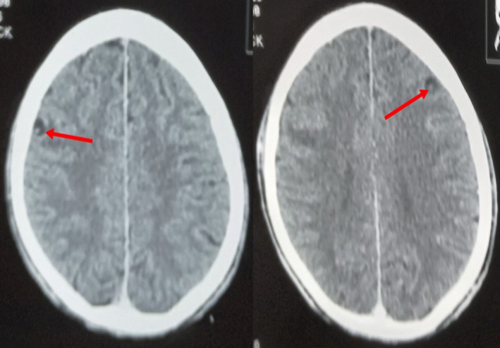
Contrast enhanced axial computed tomography (CT) scan images of the brain showing cysts with eccentric scolices in the frontal lobes.

Contrast enhanced axial computed tomography (CT) scan images of the brain showing cysts with eccentric scolices in the frontal lobes.