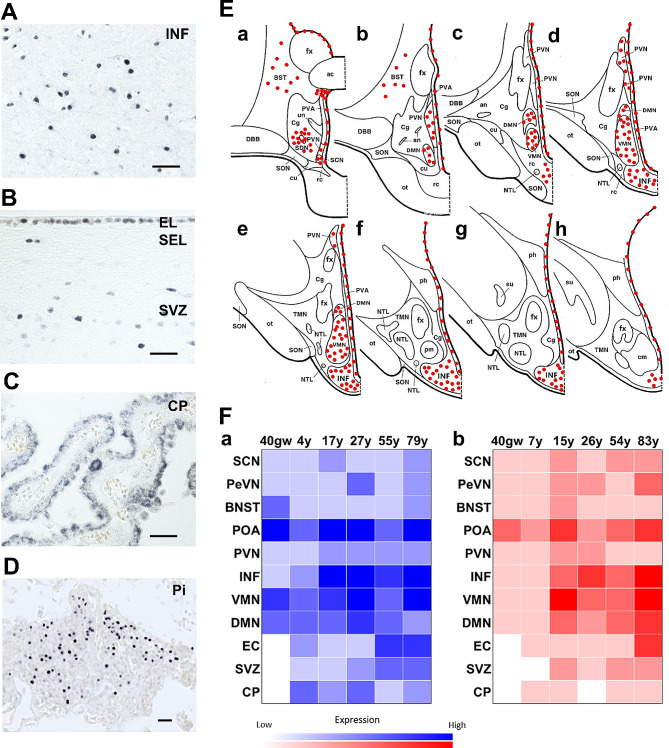
Fig. 1.
Progesterone receptor distribution in the human hypothalamus and adjacent regions. A Nuclear PR expression in the infundibular nucleus (INF). B Nuclear PR expression in cells of EL and SEL, and SVZ along lateral ventricle. C Cytoplasmic PR expression in the cuboidal and columnar epithelium of the choroid plexus. D Nuclear PR expression in the pars tuberalis (PT) of the pituitary. Scale bars: 100 μm. E Schematic illustration of PR mapping throughout the human hypothalamus. F Sex and age signature of PR-ir cells throughout human hypothalamus (N = 1). a Males (blue); b Females (red). Abbreviations: AC, anterior commissure; AN, accessory neurosecretory nucleus; BST/BNST, bed nucleus of the stria terminalis; Cg, chiasmatic gray; cm, corpus mamillare; CP, choroid plexus; cu, cuneate nucleus; DBB, nucleus of the diagonal band (of Broca); DMN, dorsomedial nucleus; EC, ependymal cell; EL, ependymal layer; FM, fasciculus mammillothalamicus; fx, fornix; INF, infundibular nucleus; GW, gestational weeks; NTL, lateral tuberal nucleus; ot, optic tract; PeVN, periventricular nucleus; ph, posterior hypothalamic nucleus; Pi, pituitary; pm, postero-medial nucleus; POA, preoptic area; PR, progesterone receptor; PVA, periventricular area; PVN, paraventricular nucleus; rc, retrochiasmatic nucleus; SCN, suprachiasmatic nucleus; SDN, sexually dimorphic (or intermediate) nucleus; SEL, subependymal layer; SON, supraoptic nucleus; su, subthalamic nucleus; SVZ, subventricular zone, TG, tuberal gray; TMN, tuberomammillary nucleus; un, unicate nucleus; VMN, ventromedial nucleus

