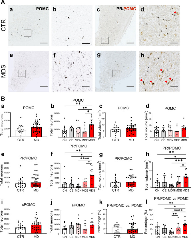
Fig. 3.
Increased numbers of PR/POMC+ neurons contribute considerably to the POMC+ neuronal increase in MDS. A POMC+ and PR/POMC+ neurons (red arrows) in the INF of a control subject and a patient with MD who died of suicide. Higher magnification of the areas framed in a, c, e and g are shown in b, d, f and h, respectively. B Analyses between CTR and MD subjects (two-group comparison), and their subsets (five-group comparison) on: total neurons and volume of POMC (a-d), PR/POMC (e-h), total neurons of sPOMC (i-j), proportion of PR/POMC neurons in POMC neurons (k-l). Note that patients with MD who died of suicides or legal euthanasia presented more PR/POMC+ neurons in the INF than patients who died naturally. Scale bars: a, c, e and g, 1 mm; b, d, f and h, 100 μm. Abbreviations: CE, control subjects who died of legal euthanasia; CN, control subjects who died of natural causes; CTR, control subjects; MD, mood disorders; MDE, subjects with mood disorders who died of legal euthanasia; MDN, subjects with mood disorders who died of natural causes; MDS, subjects with mood disorders who died of suicide; PR/POMC, neurons co-labeling progesterone receptor and pro-opiomelanocortin; sPOMC, pro-opiomelanocortin neurons that did not express progesterone receptor. Note: * indicates 0.01 ≤ P<0.05, ** indicates 0.001 ≤ P<0.01, *** indicates 0.0001 ≤ P<0.001, **** indicates 0.00001 ≤ P<0.0001, Global-P<0.05

