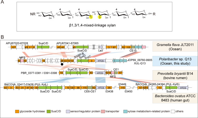
Fig 1.
The xylan utilization locus, XUL-Q13, in Polaribacter sp. Q13. (A) Schematic of MLX. The β1,3-linkage is highlighted with yellow solid circles. NR, non-reducing end; R, reducing end. (B) Synteny of XUL-Q13 and known XULs from animal gut and marine Bacteroidetes. The sequence alignment was performed with BLAST+ (version 2.7.1+, E-value 1e−5). Sequence similarities are symbolized by red for direct comparisons and blue for reversed comparisons. Darker colors correspond to higher identities. Numbers labeled in orange arrows indicate GH families. Xyn26A is marked with a solid black circle. HTCS, hybrid two-component sensor; MFS, major facilitator superfamily transporter; CE, carbohydrate esterase.

