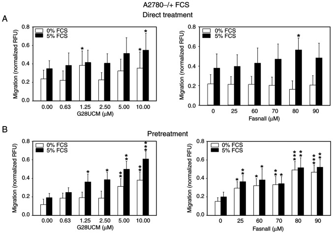
Figure 1.
Migration of OC cells is stimulated by FASN inhibitors even in the absence of serum. (A) 5×10⁴ red fluorescent mCherry-A2780 OC cells were plated in media containing 0.4% FCS in 24-well FluoroBlok cell culture inserts (apical chamber) with pores of 8 μm diameter, which were inserted into the basal chambers of 24-well plates containing media with 0% (white bars) or 5% FCS (black bars) as chemoattractant. Subsequently, solvent (DMSO, final concentration ≤0.1%) without (control groups) or with various concentrations of the FASN inhibitors, G28UCM or Fasnall (experimental groups), was added ('Direct Treatment'). (B) Fluorescent mCherry-A2780 cells were first pretreated for 24 h with plain solvent (control group) or with various concentrations of FASN inhibitors G28UCM or Fasnall in RPMI-1640 containing 5% FCS (experimental groups). Solvent- and FASN-inhibitor pretreated mCherry-A2780 OC cells were then transferred to drug-free RPMI-1640 with 0.4% FCS and plated in the apical chamber. Basal chambers contained media with 0% (white bars) or 5% FCS (black bars; 'Pretreatment'). In both experimental settings presented in A and B, fluorescent cells were then allowed to migrate for 24 h through the pores from the apical chamber down to the basal chamber before determining fluorescence in the basal chamber with a fluorometer. Data are provided in RFU normalized to seeded mCherry-A2780 cell number. All data are presented as the mean ± SD, n ≥3. Data were analyzed using two-way ANOVA, followed by a Tukey's post hoc test. *P<0.05, **P<0.01 and ***P<0.001, relative to the solvent treated control (0 μM drug) in 0% (white bars) or 5% FCS (black bars), respectively. OC, ovarian cancer; FASN, fatty acid synthase; FCS, fetal calf serum; RFU, relative fluorescence units; SD, standard deviation.

