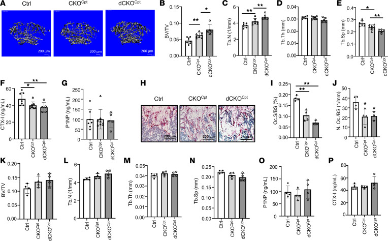
Figure 5. Deletion of Cpt1a suppresses osteoclast formation and increases bone mass in female mice only.
(A) Representative μCT images of reconstructed trabecular bone of distal femurs. (B–E) Quantification of trabecular bone parameters by μCT. (F and G) Serum CTX-I and P1NP levels. (H) Representative TRAP staining images of distal femoral sections. Scale bar: 200 μm. (I and J) Quantification of osteoclast surface (I) and number (J) on distal femoral sections. (K–N) Quantification of trabecular bone parameters by μCT in male mice with indicated genotypes. (O and P) Serum CTX-I and P1NP levels of male mice with different genotypes. *P < 0.05, **P < 0.01, against Ctrl unless otherwise indicated, 1-way ANOVA with Tukey’s multiple comparisons test. Error bars: SD.

