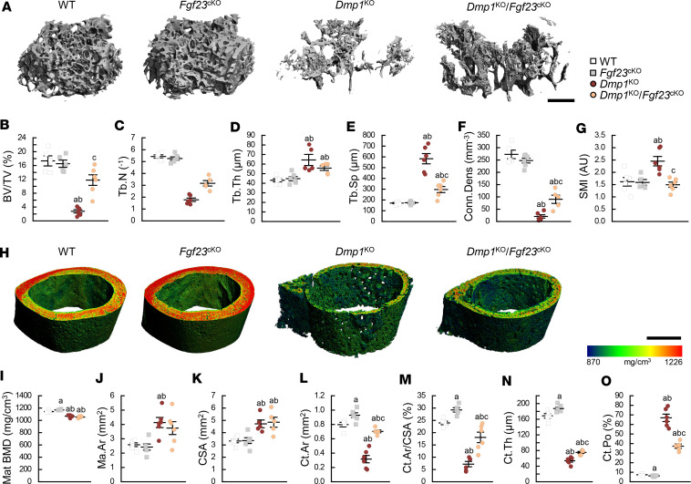
Figure 4. Osteocyte-specific deletion of Fgf23 partially restores bone microarchitecture in Dmp1KO mice.
3D μCT (A) scan reconstruction of distal femur trabecular metaphysis (scale bar = 500 μm) and (B–G) parameters of trabecular bone microarchitecture; (H) scan reconstruction of midshaft femur cortical diaphysis (scale bar = 500 μm) and (I–O) parameters of cortical bone microarchitecture. All analyses were performed in 12-week-old WT (n ≥ 5), Fgf23Dmp1-cKO (n ≥ 5), Dmp1KO (n ≥ 5), and Dmp1KO Fgf23Dmp1-cKO (n ≥ 5) mice. Values are expressed as mean ± SEM; P < 0.05 vs. aWT, bFgf23cKO, cDmp1KO. Statistical tests were ANOVA test followed by post hoc t tests and multiple-testing correction using Holm-Bonferroni method. BV/TV, bone volume to tissue volume ratio; Tb.N, trabecular number; Tb.Th, trabecular thickness; Tb.Sp, trabecular separation; Conn.Dens, connectivity density; SMI, structural model index; mat BMD, material bone mineral density; Ma.Ar, marrow area; CSA, cross-sectional area; Ct.Ar, cortical area; Ct.Th, cortical thickness; Ct.Po, cortical porosity.

