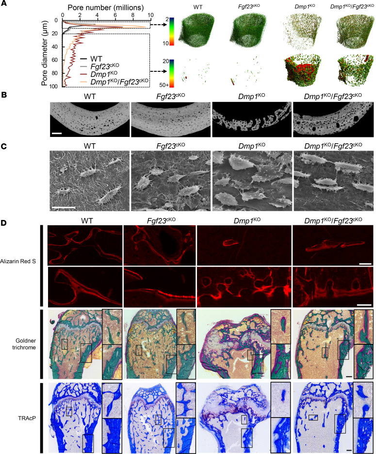
Figure 5. Osteocyte-specific deletion of Fgf23 partially restores mineralization and lacuno-canalicular network in Dmp1KO mice.
(A and B) High-resolution μCT analysis of cortical bone porosity (scale bar = 100 μm), (C) acid-etched scanning electron microscopy of femur cortical bone (scale bar = 20 μm), (D) red fluorescence microscopy imaging of ARS-stained mineralization fronts (top), and bright-field microscopy imaging of modified trichrome Goldner staining (middle) and tartrate-resistant acidic phosphatase (TRAcP) staining (bottom) of longitudinal histology sections of distal femur (scale bar = 100 μm for ARS, 250 μm for Goldner and TRAcP). For each staining, top (×3.5 original magnification) and bottom (×1.8 original magnification) zoom-in panels represent regions of interest in trabecular and in cortical bone, respectively. All analyses were performed in 12-week-old WT (n ≥ 5), Fgf23Dmp1-cKO (n ≥ 5), Dmp1KO (n ≥ 5), and Dmp1KO Fgf23Dmp1-cKO (n ≥ 5) mice.

