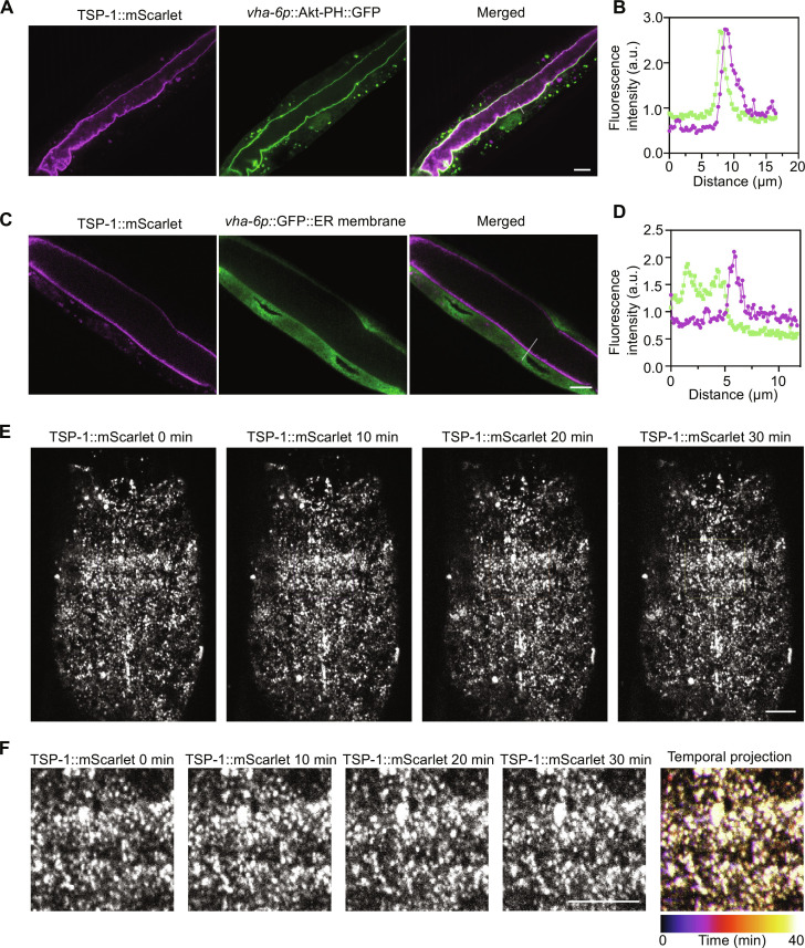
Fig. 4. Subcellular localization and stability of tetraspanin web formed by endogenous TSP-1::wrmScarlet.
(A) Representative confocal images showing colocalization of the marker Akt-PH::GFP, which binds to intestinal apical plasma membrane inner leaflet PIP2/PIP3, with endogenous TSP-1 tagged with wrmScarlet by CRISPR. Scale bars, 10 μm. (B) Fluorescent intensity correlation analysis showing close proximity of Akt-PH::GFP and TSP-1::wrmScarlet. (C) Representative confocal images showing non-colocalization of the marker ERm::GFP, which labels intestinal ER membrane, with endogenous TSP-1 tagged with wrmScarlet by CRISPR. Scale bars, 10 μm. (D) Fluorescent intensity correlation analysis for ERm::GFP and TSP-1::wrmScarlet. (E) Representative confocal time-series images showing stability of tetraspanin webs formed by endogenous TSP-1::wrmScarlet, with enlarged views in (F). Scale bars, 10 μm.

