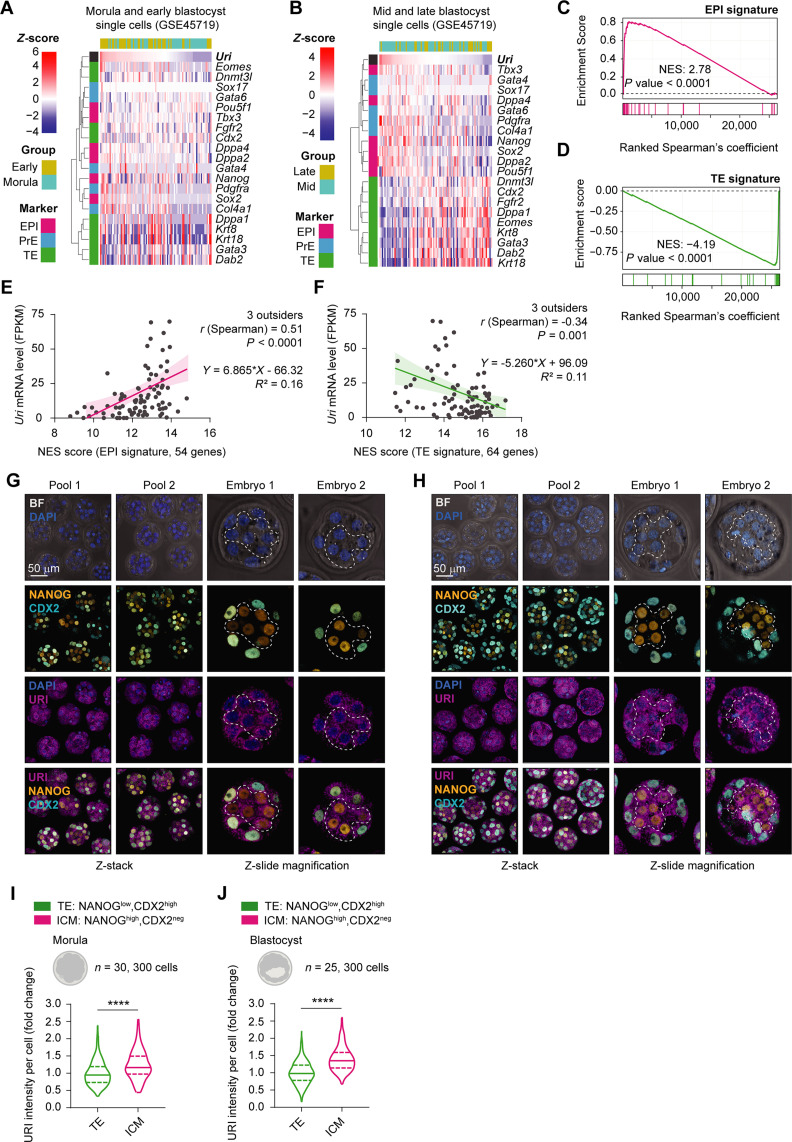
Fig. 2. High URI expression is restricted to the EPI compartment.
(A) Heatmap showing Uri-ranked mRNA expression of EPI (fuchsia), PrE (blue), and TE (green) marker genes in morula and early blastocysts from scRNA-seq datasets. (B) Heat map showing Uri-ranked mRNA expression of EPI, PrE, and TE marker genes in mid and late blastocyst single cells from referenced RNA-seq datasets. (C and D) GSEA showing EPI (C) or TE (D) gene signature distribution in ranked gene correlation with Uri expression from (A and B, respectively). (E and F) Linear regression analysis of normalized Uri mRNA levels and normalized enrichment score (NES) of GSEA for EPI (C) or TE signature (D). (G and H) IF of URI, NANOG, and CDX2 in pooled morula (G) or blastocyst embryos (H). Magnification pictures for single embryos are shown. Dashed lines delimit ICM compartment. Scale bar, 50 μm. (I and J) URI intensity in ICM and TE compartment from morula (I) or blastocyst (J) embryos from (G and H, respectively). t test; ****P < 0.0001. Total number of embryos is referred in each panel. Repository accession number for sequencing dataset analysis are indicated in respective panel and compiled in table S1. Gene signature lists are arranged in table S2.

