
Figure 1.
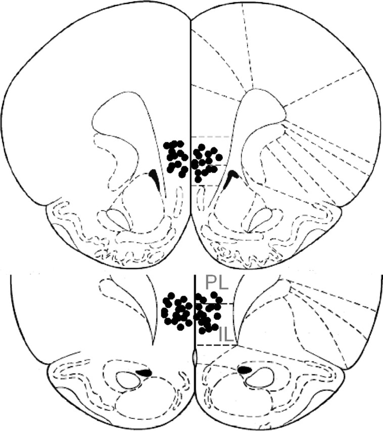
Schematic drawing of IL (infralimbic cortex) and PL (prelimbic cortex) injector tip positions (together they compose the vmPFC). Shown is a coronal view at position 3.20 and 2.70 mm anterior to bregma. Adapted with permission from Elsevier 1986, Paxinos and Watson 1986.