
Fig. 4.
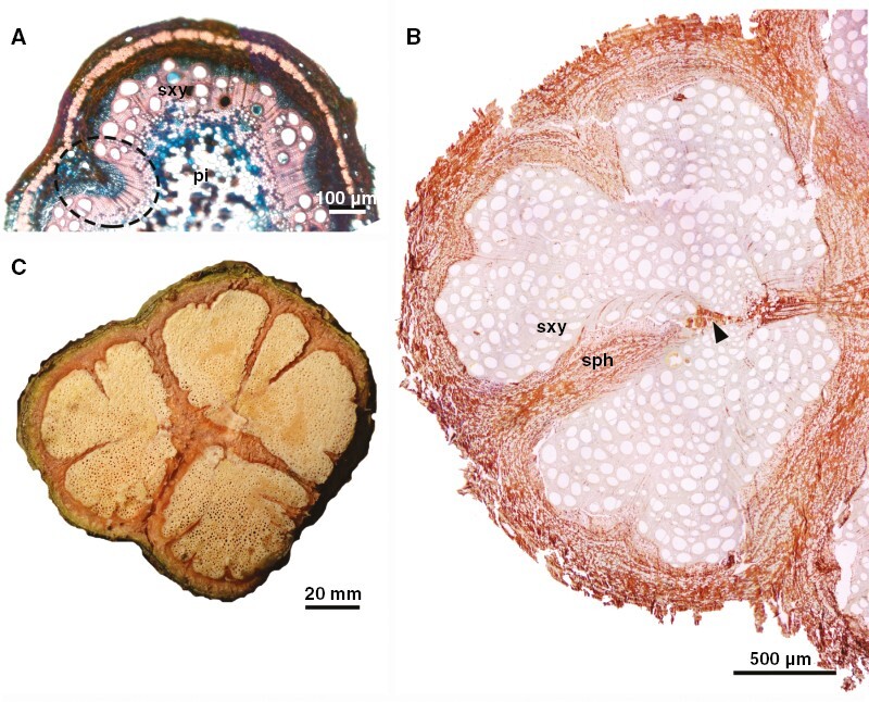
Developmental anatomy of stems of U. laevis (ontogeny 3). (A) Early stage of secondary growth showing premature formation of phloem wedges (ellipse). (B) Light microscopy (unstained) of developmental stage presented in (C), showing different sizes of phloem wedges dissecting the vascular system. Note also disruptive parenchyma (arrowhead), which also contributes to vascular tissue dissection. (C) Macroscopy of stem showing late developmental stages where furrows have fissured the vascular cylinder. sph, secondary phloem; sxy, secondary xylem; pi, pith.