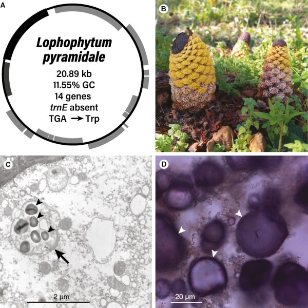
Fig. 3.

Lophophytum pyramidale. (A) Map of the plastome with the main features shown in the centre. (B) Inflorescences of L. pyramidale growing in the province of Misiones (Argentina). Yellow and whitish flowers are male and female, respectively. (C) Transmission electron micrograph of an ovarian cell. The arrow points to the non-photosynthetic plastid. (D) Light micrograph of a cell from the female flower stained with iodine. Arrowheads depict starch granules.
