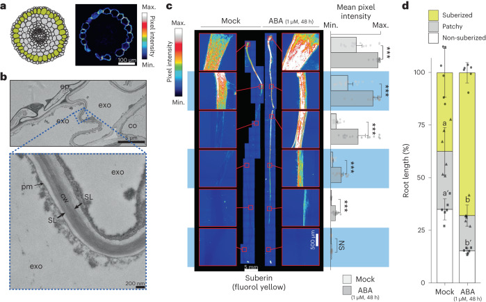
Fig. 1. Suberin is deposited in the tomato exodermis and is regulated by ABA.
a, Graphical representation (left) of S. lycopersicum (cv. M82) root anatomy (the exodermis is highlighted in yellow) and representative cross-section (right) of a 7-day-old root stained with FY. Scale bar, 100 µm. b, Transmission electron microscopy cross-sections of 7-day-old roots obtained at 1 mm from the root–hypocotyl junction. Top: the epidermal (ep), exodermal (exo) and inner cortex (co) layers. Bottom: a close-up of the featured region (zone defined with blue dotted lines), showing the presence of suberin lamellae (SL). cw, cell wall; pm, plasma membrane. c, Fluorol yellow (FY) staining for suberin in wild-type 7-day-old plants treated with mock or 1 µM ABA for 48 h. Whole-mount staining of primary root (left) and mean fluorol yellow signal along the root (right), n = 6; error bars, s.d. Asterisks indicate significance with one-way analysis of variance (ANOVA) followed by a Tukey-Kramer post hoc test (***P < 0.005). NS, not significant. d, Developmental stages of suberin deposition of wild-type plants treated with mock or 1 µM ABA for 48 h. Zones were classified as non-suberized (white), patchy suberized (grey) and continuously suberized (yellow); letters indicate statistically different groups; apostrophes indicate different statistical comparisons; n = 6; error bars, s.d.

