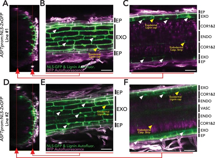
Extended Data Fig. 5. The SlASFT promoter drives exodermal-specific expression in roots.
Representative images depicting expression of the SlASFT promoter driving nuclear localized GFP (SlASFTp:NLS-2xGFP) in two independently-transformed hairy root lines. NLS-GFP and lignin autofluorescence (Green) and epidermal RFP autofluorescence (Magenta). (A and D) Transversal z-stack projections of mature regions of transgenic hairy roots. Red lines indicate the planes shown in the subsequent longitudinal sections. (B and E) Longitudinal section of a top plane of the z-stacks, showing the epidermal and exodermal cell layers. White arrows indicate some of the GFP-tagged nuclei in the exodermis. Yellow arrows indicate the lignin autofluorescence of the exodermal polar cap. (C and F) Longitudinal section of a bottom plane of the z-stacks, showing the epidermis, exodermis, cortex layers 1 and 2, and endodermis. White arrows indicate the GFP-tagged nuclei in the exodermis. Yellow arrows indicate the lignin autofluorescence of the exodermal polar cap and the endodermal Casparian Strip. EP: Epidermis; EXO: Exodermis; COR1&2: Cortex layer 1 and 2; ENDO: Endodermis, VASC: Vasculature.

