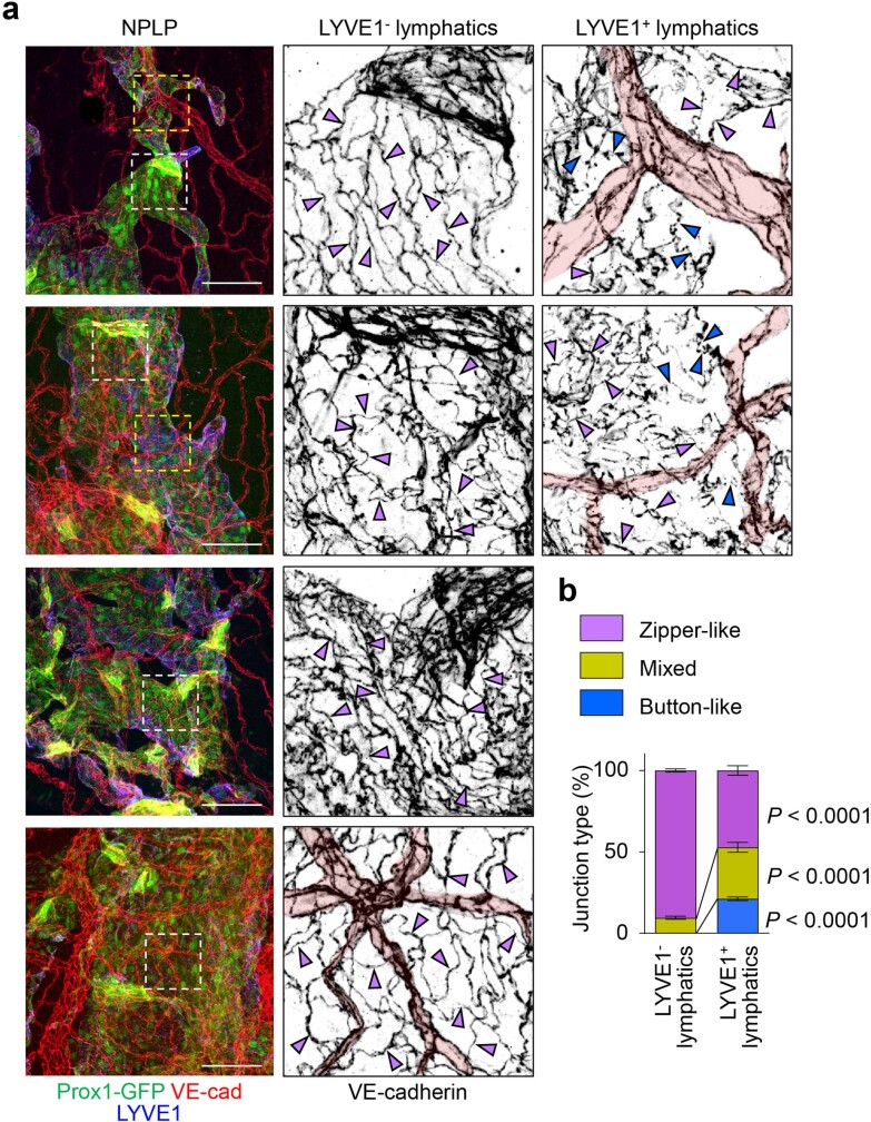
Extended Data Fig. 3. Intercellular junctions of lymphatic endothelial cells of the nasopharyngeal lymphatic plexus.
a,b, Immunofluorescence images of whole mounts of the dorsal and ventral portions of nasopharyngeal lymphatic plexus (NPLP) stained for VE-cadherin (VE-cad) and LYVE1 in adult (8–10 weeks old) Prox1-GFP mice. White and yellow dashed-line box areas are enlarged in the centre and right panels. Blood capillaries are highlighted in pink. Zipper-like intercellular junction (magenta arrowheads) predominated (95%) in lymphatics with little or no LYVE1 staining, whereas button-like intercellular junctions (blue arrowheads) were more numerous (21%) in LYVE1+ lymphatics. Scale bars, 100 μm. Similar findings were obtained from n = 4 mice in two independent experiments. Bars indicate mean ± s.e.m. P values for junctions in LYVE1- and LYVE1+ lymphatics were calculated by two-way ANOVA test followed by two-tailed Holm-Sidak’s multiple comparison post-hoc test.

