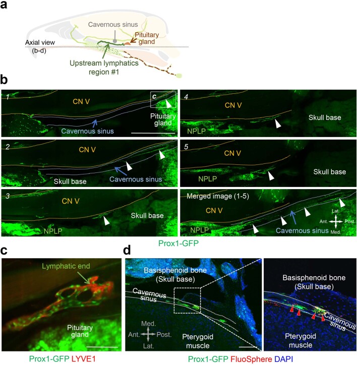
Extended Data Fig. 4. Prox1+/LYVE1+ lymphatics near the pituitary gland extend to the nasopharyngeal lymphatic plexus along cranial nerve V and cavernous sinus of Prox1-GFP mice.
a, Diagram of a sagittal view of dural lymphatics designated upstream lymphatics region #1. b, Light-sheet fluorescence microscopic images showing serial optical sections (numbered) of upstream lymphatics region #1 (white arrowheads) originating near the Prox1+ pituitary gland. These lymphatics course along the cavernous sinus (outlined with blue-dotted lines) and beneath cranial nerve (CN) V (outlined with yellow-dotted lines) en route to the nasopharyngeal lymphatic plexus (NPLP). Anatomical positions are indicated in the lower right corner. Scale bar, 1 mm. Similar findings were obtained from n = 5 mice in three independent experiments. Ant., anterior; Post., posterior; Med., medial; Lat., lateral anatomical position. c, Light-sheet fluorescence microscopic image showing LYVE1-stained (red), blunt-ended Prox1+/LYVE1+ dural lymphatics (green nuclei) in an enlargement of the region in b section 1 marked by white-dotted line box (c) near the Prox1+ pituitary gland (bright green). Scale bar, 100 μm. d, Fluorescence microscopic images of section showing Prox1+ upstream lymphatics region #1 containing FluoSpheres (red arrowheads) along the cavernous sinus. The region of the white dashed-lined box is enlarged in the right panel. Anatomical positions are indicated in the lower left corner. Scale bar, 200 μm. Similar findings were obtained from n = 3 mice in two independent experiments. Ant., anterior; Post., posterior; Med., medial; Lat., lateral anatomical position.

