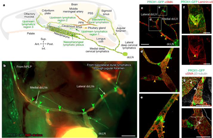
Fig. 3. Connections of the NPLP and features of deep cervical lymphatics.
a, Diagram of intracranial upstream lymphatic regions 1, 2 and 3, which drain through the NPLP en route to medial deep cervical lymphatics and dcLNs in the neck. Upstream lymphatic region 1 includes the lymphatics near the pituitary gland and cavernous sinus that drain to the NPLP. Upstream lymphatic region 2 includes the lymphatics in the anterior region of basolateral dura near the middle meningeal artery and petrosquamosal sinus (PSS) that course along the PPA to the NPLP. Upstream lymphatic region 3 includes lymphatics near the cribriform plate that drain to the lymphatics in the olfactory mucosa en route to the posterior nasal lymphatic plexus and NPLP. By contrast, the lymphatics in the posterior region of the basolateral dura around the sigmoid sinus do not drain to the NPLP but, instead, pass through the jugular foramen to lateral deep cervical lymphatics en route to dcLNs. Anatomical positions are indicated at the bottom left. b, Fluorescence image showing medial dcLVs, lateral dcLVs, lymphatic valves (green arrowheads) and TMR–dextran (red) in lymphatics deep in the neck of a Prox1-GFP mouse. The image was obtained 30 min after i.c. infusion of TMR–dextran (molecular mass, 10 kDa) at 1.0 μl min−1 for 3 min. Medial dcLVs connect to the NPLP, and lateral dcLVs connect to the basolateral dural lymphatics through the jugular foramen. Scale bar, 1 mm. Similar findings were obtained from n = 6 mice in three independent experiments. c–e, Immunofluorescence images of whole mounts showing the distributions of PROX1-dense, semi-lunar shaped lymphatic valves (green arrowheads) and αSMA+ circular smooth muscle cells (SMCs, orange arrowheads) in the medial and lateral dcLVs. d,e, Magnified images of the regions indicated by the green boxes in c. Scale bar, 1 mm (c). Similar findings were obtained from n = 4 mice in two independent experiments. f, Immunofluorescence images of whole mounts showing a typical semi-lunar-shaped PROX1-dense, laminin-α5high valve (yellow arrowheads) in a medial dcLV of a Prox1-GFP mouse. Scale bars, 200 μm. Similar findings were obtained from n = 4 mice in two independent experiments. g, Immunofluorescence images of whole mounts showing the distributions of β3-tubulin+ axons (white arrowheads) and αSMA+ circular smooth-muscle cells (red) along dcLVs. Scale bars, 200 μm. Similar findings were obtained from n = 4 mice in two independent experiments.

