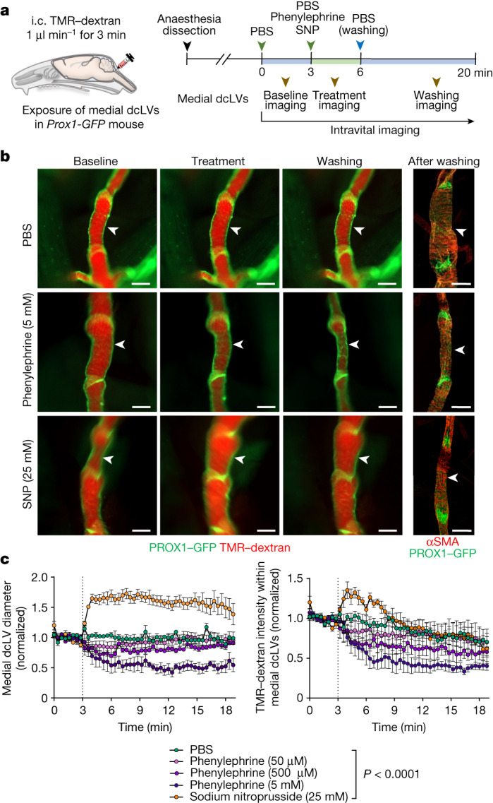
Fig. 5. Regulation of CSF outflow by myogenic control of medial deep cervical lymphatics.

a, Diagram of the experimental sequence for intracisternal infusion of TMR–dextran at 1.0 μl min−1 for 3 min, and intravital imaging of medial deep cervical lymphatics (medial dcLVs) during pharmacological manipulation in Prox1-GFP mice. SNP, sodium nitroprusside. b, Fluorescence images showing TMR–dextran fluorescence in medial dcLVs (white arrowheads) at 2 min before treatment, during treatment and during washing. Right, immunofluorescence images of whole mounts stained for PROX1-dense lymphatic valves and αSMA+ circumferential smooth-muscle cells in medial dcLVs at 10 min after washing. Scale bars, 200 μm. Similar findings were obtained from n = 4 mice in three independent experiments. c, Changes in diameter and TMR–dextran fluorescence in the medial dcLVs over 17 min after the onset of five different pharmacological manipulations (vertical dotted lines). Data are mean ± s.e.m. n = 4 mice per group from four independent experiments. Values were normalized to the mean baseline value for each group. P values were calculated using two-way repeated-measures ANOVA.
