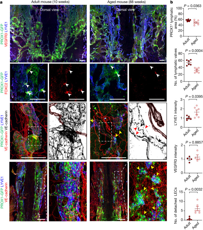
Fig. 6. Ageing-related alterations in the NPLP.
a, Immunofluorescence images of whole mounts showing the dorsal surface of the NPLP in adult (aged 10 weeks) and aged (aged 88 weeks) mice. Multiple abnormalities are evident in the aged mice. Row 1, the lymphatic plexus is smaller and has fewer valves. Row 2, PROX1-dense, FOXC2+ lymphatic valves (white arrowheads) are less numerous. Rows 3 and 4, the lymphatic plexus is smaller (the regions indicated by white dashed boxes are magnified in the adjacent monochrome panels); fewer LECs have an oak-leaf shape (black arrowheads); LECs have altered intercellular junctions (red arrowheads); and some cells appear to be detached from the adjacent cells (yellow arrowheads). The blood capillaries are marked by a pink overlay. Scale bars, 200 μm. Similar findings were obtained from n = 7 mice in three independent experiments. b, Comparison of the lymphatic area, valves, LYVE1 and VEGFR3 staining, and detached endothelial cells in the plexus between adult (aged 8–12 weeks) and aged (aged 73–102 weeks) mice. Each dot is the value for one mouse; n = 7 (PROX1+ lymphatic area), n = 6 (number of lymphatic valves), n = 6 (LYVE1 intensity), n = 4 (VEGFR3 intensity) and n = 6 (number of detached LEC) mice per group from three independent experiments. Data are mean ± s.e.m. P values were calculated using two-tailed unpaired t-tests with Welch’s correction or two-tailed Mann–Whitney U-tests.

