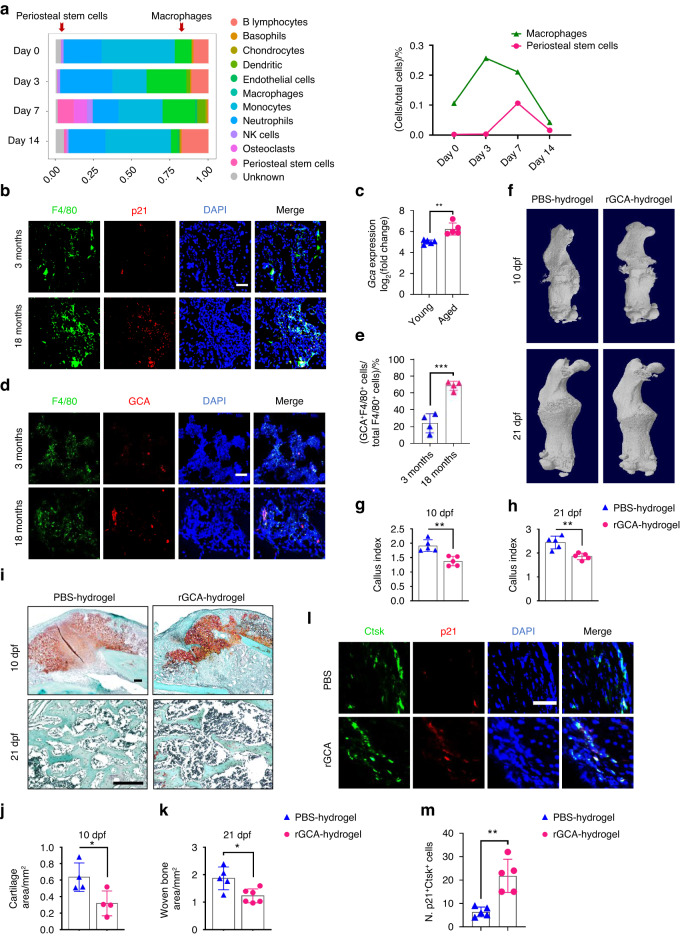
Fig. 2.
Aged macrophages in fracture calluses release GCA and impair bone regeneration. a Stacked bar chart showing the percentages of each cell type within callus tissue quantified on Day 0, Day 3, Day 7, and Day 14 post-fracture based on UMAP distribution. b Representative IF images of fracture calluses immunostained with F4/80 (green) and p21 (red) antibodies and counterstained with DAPI (blue) (n = 4). Scale bars indicate 100 µm. c Bioinformatics analysis of Gca gene expression levels in calluses from 5-month-old and 25-month-old mice at 5 days post-fracture (n = 5). d Representative IF images of fracture calluses immunostained with F4/80 (green) and GCA (red) antibodies and counterstained with DAPI (blue) (n = 4). Scale bars indicate 100 µm. e Quantification of GCA+F4/80+ cells (n = 4). f Representative micro-CT images of fractured femurs from PBS- and rGCA-treated mice at 10 dpf and 21 dpf. g, h The callus index of fractured femurs from PBS- and rGCA-treated mice at 10 dpf and 21 dpf (n = 5). i–k Safranin O staining showed cartilage callus formation and woven bone area from PBS- and rGCA-treated mouse fractured femurs at 10 dpf and 21 dpf (n = 4–6). Scale bar indicates 100 μm. l Representative IF images in the periosteum of the fracture callus at 10 days post-fracture, immunostained with Ctsk (green) and p21 (red) and counterstained with DAPI (blue). Scale bars indicate 100 µm. m Quantification of the number of p21- and Ctsk-positive cells per mm2 tissue area (N. p21+Ctsk+ cells) by Welch’s t test (n = 5). Data are presented as the means ± SDs. Unpaired t test, *P < 0.05 and **P < 0.01

