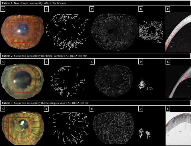
Figure 1.
Comparison of slit-lamp photography and AS-OCTA imaging for corneal vascularization. Examples of eyes (respective image rows) with corneal vascularization imaged with slit-lamp photography (panels A: visualized corneal area delineated with Fiji, see Methods, region of interest imaged by AS-OCTA highlighted with a red rectangle); panels B: segmentation of corneal vascularization using the morphological hessian-based method; panels C: segmentation of corneal vascularization using Gabor filtering), and 3 × 3-mm AS-OCTA (panels D: AS-OCTA en-face scan after post-processing in Fiji: capillary flow area delineated in white; panels E: AS-OCTA cross-sectional scan showing a vessel flow signal in red with its marked (blue) deepest extension in relation to total corneal thickness).

