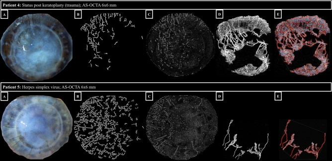
Figure 2.
Vessel segmentation using Angiotool software. The rows represent the respective patient cases. Panels A show slit-lamp photographs, panels B show segmentation of corneal vascularization (morphological hessian-based method on slit-lamp photographs), panels C show segmentation of corneal vascularization (Gabor filtering on slit-lamp photographs), panels D native OCTA 6 × 6-mm en-face scans (effectively covering 12 × 12 mm on the corneal surface, see Methods) with the capillary flow area in white, and panels E OCTA 6 × 6-mm en-face scans after Angiotool software segmentation with delineated vessels (red), and vessel branching/end points (blue). See the attached Table 1 below for corresponding vessel parameters of patient 4 and 5 (ID 4 and 5, respectively). In patient 5, the vessels in the upper sectors of the cornea are not captured by the OCTA algorithm, which may be due to slow erythrocyte flow.

