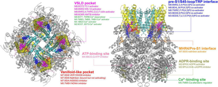
FIGURE 2.
Ligand binding sites in TRPM channels. The TRPM8 structure with TC-I 2014-bound (PDB: 6O72) is used for visualization, with the pore region, the VSLD region and cytosolic domain colored in cyan, yellow, and gray, respectively. The major binding pockets or interfaces are highlighted using different colors. The top and side views are rendered in the same way as in the TRPV channel (see Figure 1).

