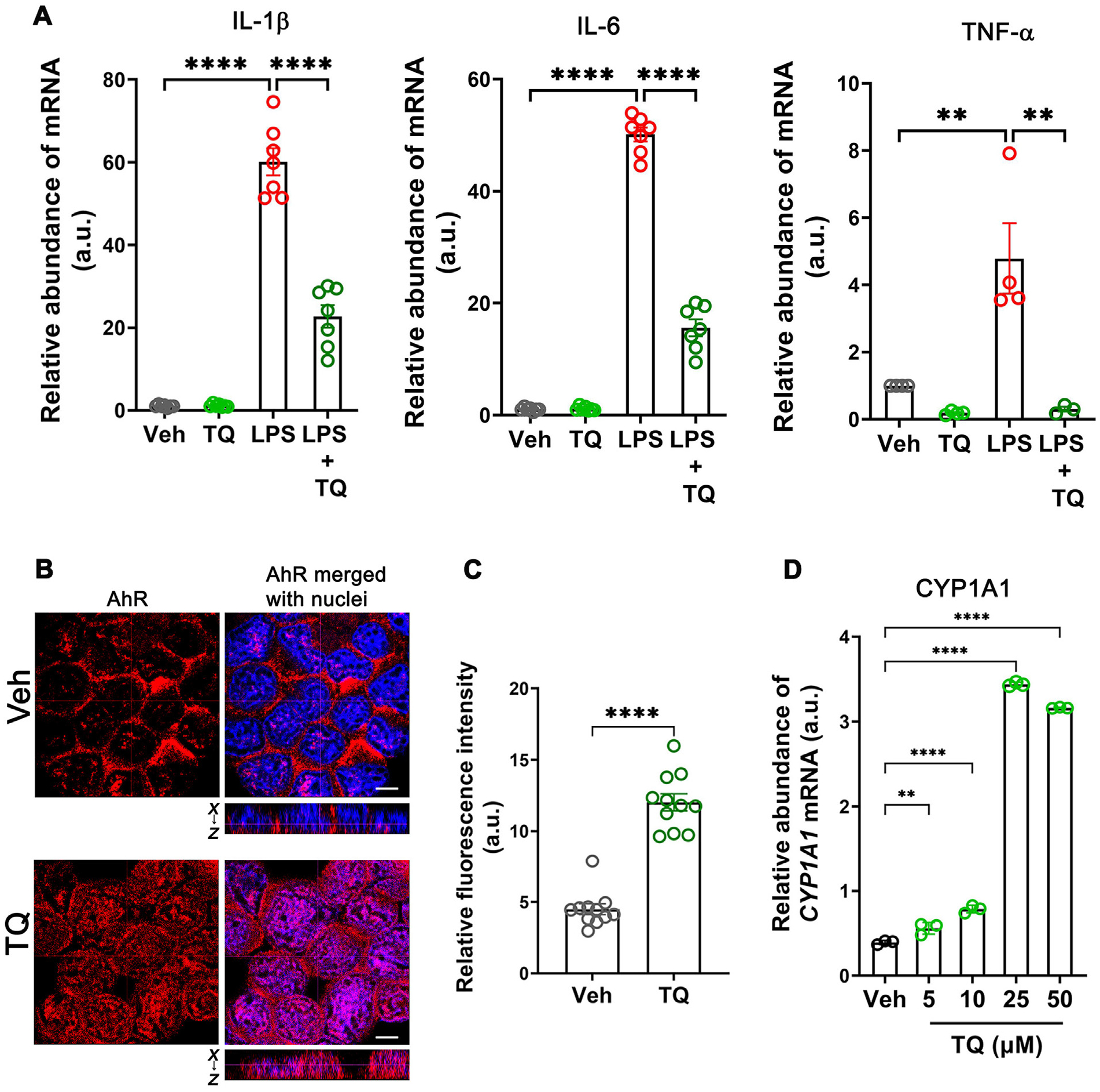
Fig. 4.

TQ exerts its anti-inflammatory effect by activating the AhR in U937 cells. (A) TQ prevented the LPS-induced (10 ng/ml) increase in the mRNA levels of IL-1β, IL-6, and TNF-α in the human U937 cell line. (B) Confocal images of AhR (red) and nuclei (blue) in U937 cells. The images showed that TQ treatment (4 h) increased AhR levels as well as its nuclear translocation, indicating that TQ activates AhR. The dotted lines represent the optical level for the x-y and x-z planes. White bar: 5 μm. These data are representative of multiple areas from independent samples, n = 5. (C) Quantification of AhR fluorescence in the nucleus from (B). (D) The mRNA levels of CYP1A1, the prototypic transcriptional target of AhR, showed a dose-dependent increase in abundance with increasing concentrations of TQ with the maximum effect observed with 25 μM concentration of TQ. Data represented as mean ± SEM, n > 3 from 3 independent experiments. Student’s t-test (C) and one-way analysis of variance with Tukey’s post test (A and D). **p < 0.01, ****p < 0.0001.
