Fig. 7.

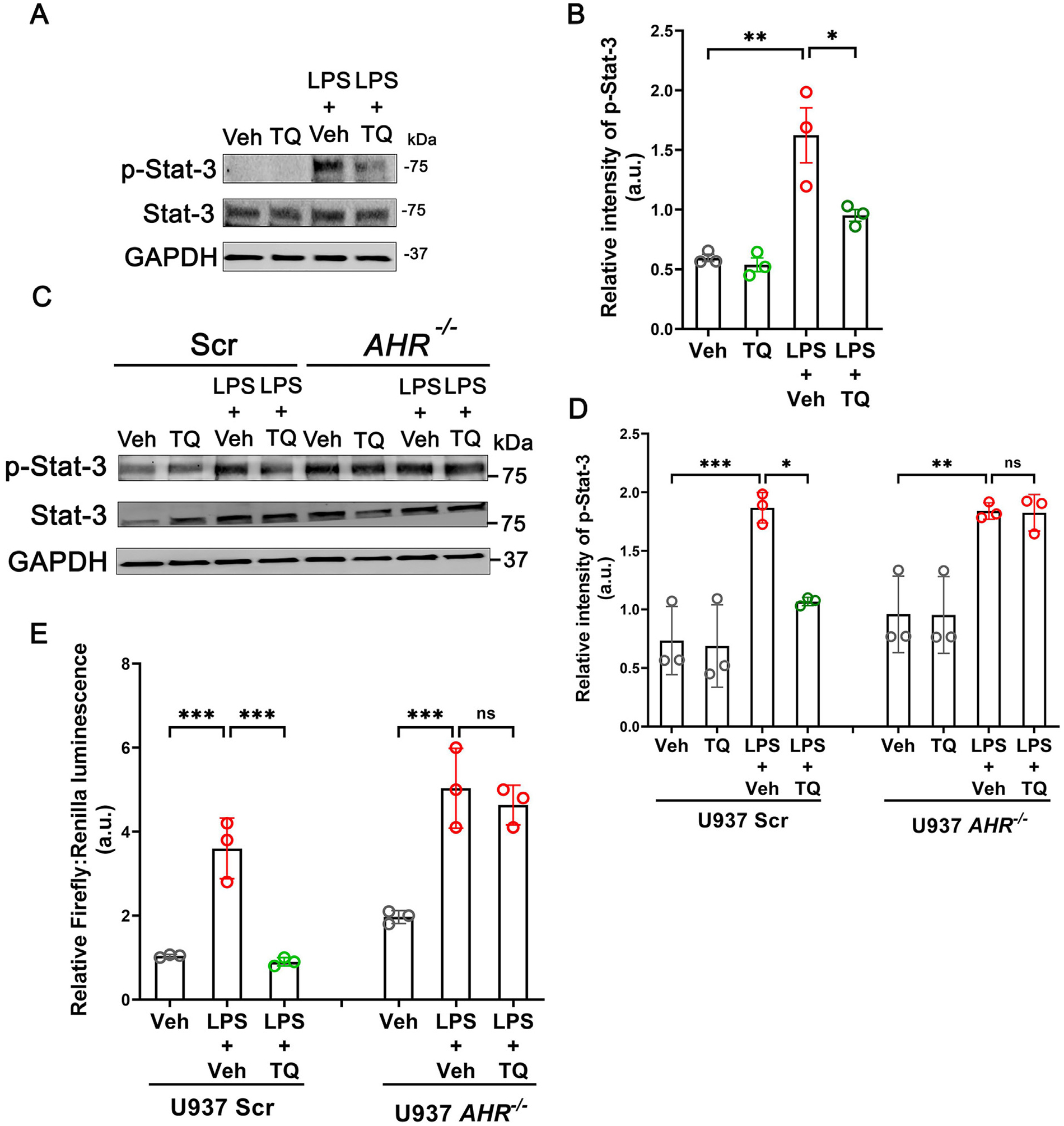
TQ reduces Stat-3 signaling. (A) TQ reduces the LPS-induced phosphorylation and activation of Stat-3 in U937 cells. The GAPDH levels are shown as the loading control. Representation of three independent experiments. (B) Quantification of p-Stat-3 band intensity normalized to total Stat-3 from (A). (C) The inhibitory effect of TQ on Stat-3 activation was dependent on AhR as AHR−/− U937 cells failed to reduce the LPS-induced phosphorylation of Stat-3 upon incubation with TQ. The GAPDH levels are shown as the loading control. Representation of three blots. (D) Densitometric quantification of phospho-Stat-3 bands normalized to total Stat-3 from (C). (E) TQ failed to prevent the LPS-induced upregulation of luciferase activity in the AHR−/− U937 cells where the luciferase reporter was placed downstream of the Stat-3 dimer binding site. Representation of 3 independent experiments. Data represented as mean ± SEM. One-way analysis of variance with Tukey’s post test. * p < 0.05, ** p < 0.01, *** p < 0.001. ns = not significant.
