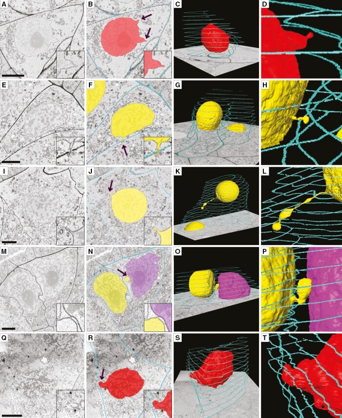
Fig. 4.

Dynamics of NP formation in meiocytes imaged by SBF-SEM. Nuclear protuberances emerge but do not cross the cell wall at the premeiotic stage (A–D). At leptotene can be seen tiny NPs (E–H), extended NPs comparable in length to the size of a nucleus (I–L) and NPs forming nucleus–nucleus contacts (M–P). At zygotene NPs that do not penetrate the cell wall appear again (Q–T). The left column presents original images, and to the right the same image is shown with cell walls and nuclei highlighted in various colours, and the two right-hand columns are 3-D reconstructions of the scanned cells and NPs with enlargement. Arrows denote NPs. Yellow and purple indicate nuclei involved in INM, red non-involved nuclei, and turquoise the outer layer of cell walls, labelled on every 25th slice. Scale bars = 5 µm.
