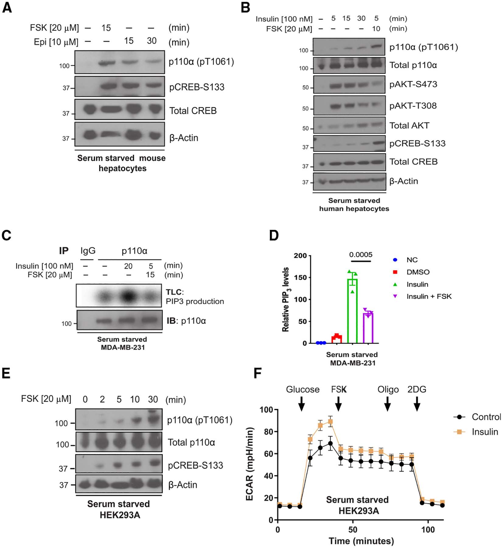
Figure 4. Forskolin and epinephrine exposure phosphorylate and inhibit p110α in cells.

(A) Immunoblot for the indicated proteins using lysates from primary mouse hepatocytes that were serum starved 12 h before stimulation with forskolin (FSK; 20 μM) for 15 min or epinephrine (Epi, 10 μM) for 15 and 30 min.
(B) Immunoblot for the indicated proteins using lysates from primary human hepatocytes that were serum starved for 12 h and treated with insulin (100 nM) for different time points (0, 5, 15, or 30 min) or insulin followed by FSK (20 μM) for 10 min.
(C) Top: radioautograph of a TLC separation demonstrating PIP3 production of endogenous p110α that was immunoprecipitated from serum-starved MDA-MB-231 cells treated with vehicle, insulin (100 nM), or insulin plus FSK (20 μM) for 15 min. Bottom: Corresponding immunoblot for p110α using the same immunoprecipitate lysate.
(D) Quantification of the radioautograph from
(C) averaged over three independent experiments. Means ± SEM. Comparisons made using ANOVA with Tukey’s multiple comparisons post-test (N = 3).
(E) Immunoblot for the indicated proteins using lysates from HEK293A cells that were serum starved for 2 h before being stimulated with FSK 20 μM for 0, 2, 5, 10, and 30 min.
(F) The extracellular acidification rate (ECAR) was monitored in serum-starved HEK293A cells with or without insulin (0.1 mM] pre-treatment for 1 h. Arrows indicate injection of glucose (10 mM), FSK (20 μM), oligomycin (Oligo; 1 μM), and 2-deoxy-D-glucose (2DG; 50 mM). Means ± SEM, N = 19 wells. Results are representative of 3 independent experiments.
