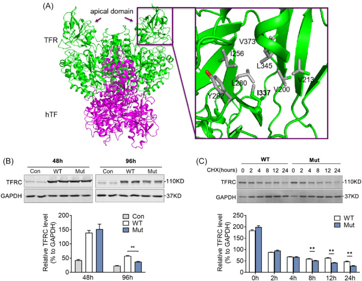
FIGURE 4.

The association of transferrin receptor (TFRC) missense variant with the instability of TFRC protein. (A) The human serum transferrin (hTF) and transferrin receptor (hTFRC) are shown as purple and green, respectively. The mutation p.I337V in apical domain and the surrounded residues are shown as sticks and labeled. Hydrophobic groups in the surrounded residues are colored gray. The Cα group of p.I337 is shown as sphere, and the Cδ group of p.I337 is shown as gray stick. Residues 339–343 are removed for clarity. This figure was prepared with PyMOL using PDB 3S9M. (B) Western blot analysis and quantification of the TFRC protein levels of wild‐type (WT) and mutant (Mut) TFRC in HEK293T cells after transfection at indicated time. (C) HEK293T cells after transfection with WT and Mut TFRC were treated with 50 μg/mL cycloheximide (CHX) at indicated time and the TFRC protein levels were detected by western blot and quantified. Data were shown as mean ± SEM and *p < .05, **p < .01 compared to control group. Con, controls.
