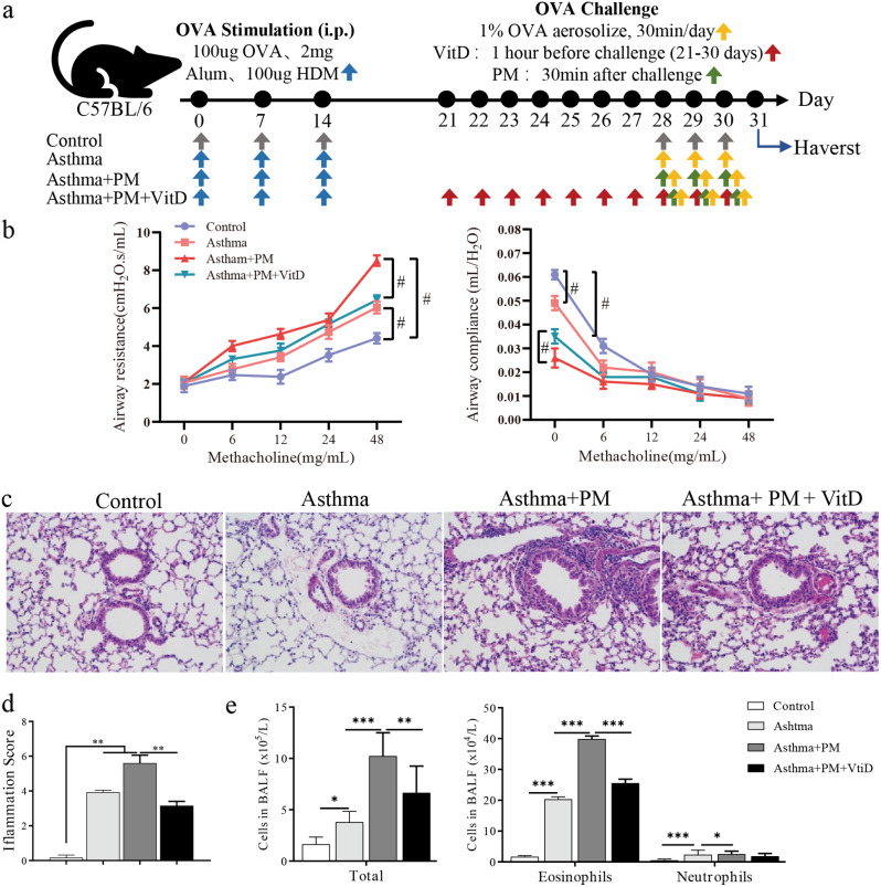
Fig. 1.
Vitamin D improves PM-exposed lung histopathological inflammation in asthmatic mice. Thirty-two female mice were randomly assigned to the Control group, Asthma group, Asthma + PM group, or Asthma + PM + Vit D group. (a) Schematic illustration of the mouse modeling process. The gray arrows indicate that each mouse was administered normal saline instead. The blue arrow indicates that each mouse was injected intraperitoneally with OVA sensitization solution. The yellow arrow indicates that the mice were exposed to 1% OVA for 30 min. The green arrow indicates that the mouse was intratracheally instilled with 100ug of PM. The red arrow indicates that the mouse was gavaged with Vit D. (b) Airways resistance (Rrs) and compliance (Crs) of different groups (#p < 0.01). (c) Airway inflammation (H&E staining, ×200). (d) Inflammatory index. (e) proportion of inflammatory cells in BALF (* p < 0.05, ** p < 0.01, *** p < 0.001, and **** p < 0.0001)

