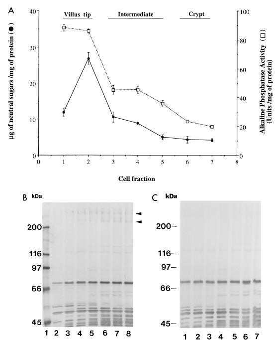
FIG. 3.
Distribution of IMTGP-1 and IMTGP-2 along the crypt-villus axis. Intestinal epithelial cells were sequentially eluted as described in Materials and Methods. (A) The eluted cell fractions were assayed for neutral sugar content (•) and alkaline phosphatase activity (□). Glycoproteins (50 μg) from each cell fraction were separated by SDS-PAGE (7% polyacrylamide) and electrotransferred to a PVDF membrane. Fractions 1 to 7 in panel A correspond to lanes 2 to 8 of adhesive phenotype (B) and lanes 1 to 7 of nonadhesive phenotype (C). K88ac adhesin binding activity was detected with biotinylated K88ac adhesin.

