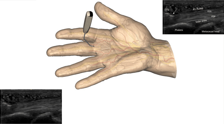
Figure 7. Beginning of the A1 pulley release by using the TF-SI.
Note the cutting part of the instrument, represented by a straight hyperechoic line on the sonographic image (arrow), thanks to its specific flange. To better understand the sonographic view, the image on the left and bottom is repeated right at the top with a legend. (FDS: Flexor digitorum superficialis; FDP: Flexor digitorum profundus) Note that the sonography probe is intentionally not shown; better observe the cutting instrument underneath the skin. The probe is used throughout the procedure.
Image created by F Moungondo.

