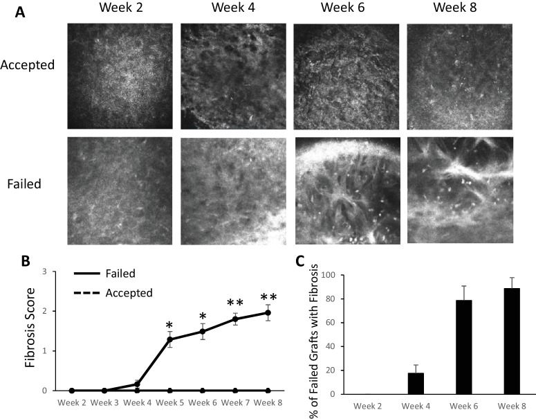
Figure 2.
In vivo confocal microscopy evaluation of fibrosis in corneal transplantation. (A) Representative in vivo confocal microscopy images showing the development of hyperreflective, thickened bands in the anterior stroma in failed grafts (bottom row). (B) Fibrosis score increased in failed grafts (solid line) over time, but accepted grafts continued to have a corneal opacity score of 0 (dashed line). (C) The majority of failed grafts demonstrated signs of fibrosis on confocal microscopy, and fibrosis became most evident around week 6. *P < 0.05, **P < 0.005 (failed, n = 16; accepted, n = 14).

