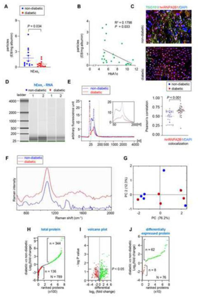
Figure 4: Human chronic wound fluid of diabetic subject has a low abundance of Keratinocyte-derived exosomes with compromised cargo loading:
A, Quantification of from chronic non-diabetic and diabetic human wound fluid normalized with albumin. B, Regression analysis of the abundance of keratinocyte-originated exosomes with HbA1C. C, Representative fluorescence images of human non-diabetic and diabetic wound-edge epidermis showing immunofluorescence staining of hnRNPA2B1(red) and TSG101 (green) with DAPI counterstaining in. Scale, 50 μm. D, High-resolution automated electrophoresis of RNA in isolated from human chronic wound fluid of non-diabetic and diabetic subjects shows more abundance of RNA in non-diabetic human wound fluid compared to diabetic human wound fluid. E, Comparison of bioanalyzer generated electropherograms of RNA in isolated from human chronic wound fluid of non-diabetic and diabetic subjects. F, Representative Raman spectra of isolated from non-diabetic and diabetic chronic wound fluid. G, 2D-score plot constructed from principal component analysis of Raman spectra. H, Ranked protein plot showing intensity of proteins in isolated from human chronic wound fluid of non-diabetic and diabetic subjects. I, Volcano plot showing proteins with a p value <0.05 are above the threshold line. Note that only significant proteins with a p < 0.05 have a -log10(.05) > 1.3. J, Ranked plot of the proteins that were significantly different between t isolated from human chronic wound fluid of nondiabetic and diabetic subjects. Data in A and C were shown as mean ± SEM and analyzed by Student’s t-test.

