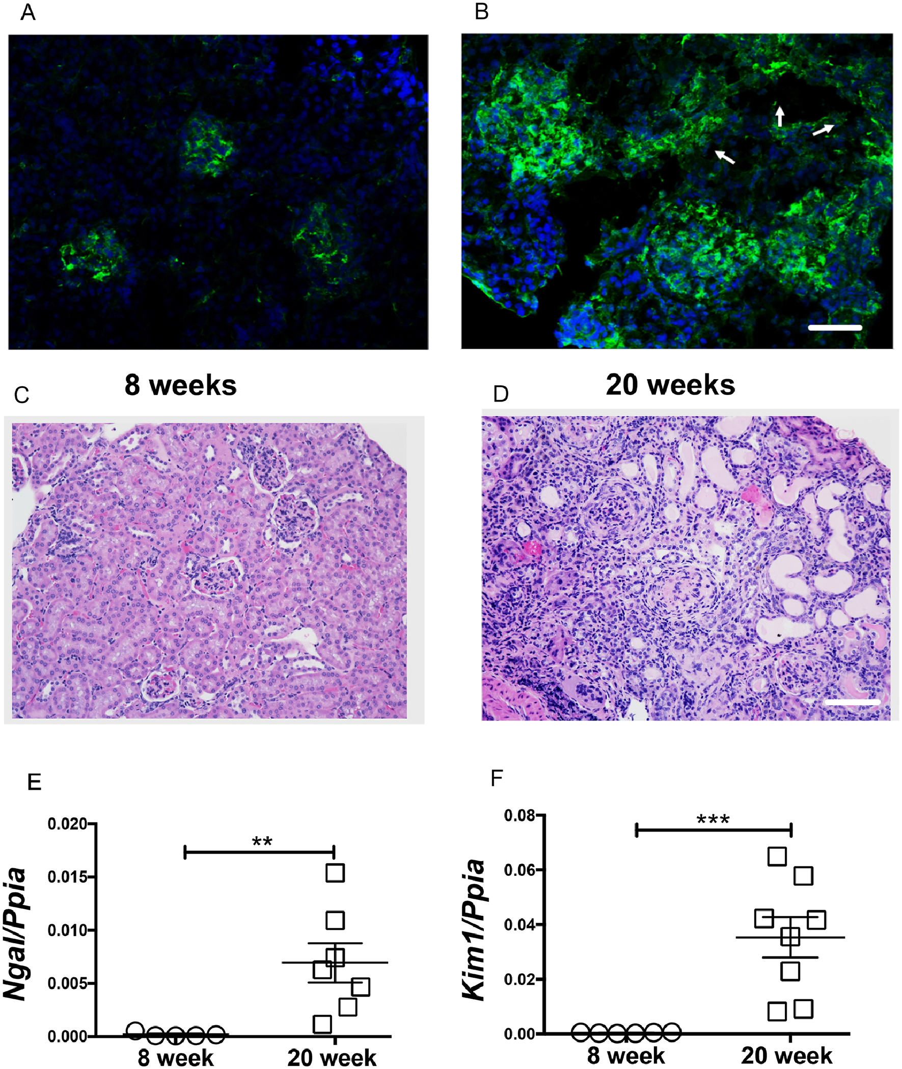
Figure 1. Tubular injury is a distinct feature in lupus nephritis.

Glomerular immune complexes were observed in 8-week-old non nephritic female MRL/lpr mice (A). At 20-weeks of age, glomerular and tubular immune complex deposits are evident (arrow points to the tubules) (B). Compared to 8-week-old female (C), the renal histology (H&E) at 20 weeks showed severe periglomerular and interstitial immune infiltrates. Along with the traditional glomerular injury, large number of a-nuclear tubules, tubular cast are also evident (D). Scale bar = 50 μm and 100 μm. The tubular injury marker Ngal and Kim1 were significantly elevated in 20-week-old nephritic mice (E-F). Statistical significance was determined by 2-tailed Mann-Whitney test and represented as mean ± SEM. **P < 0.01, ***P < 0.001.
