Fig. 1.
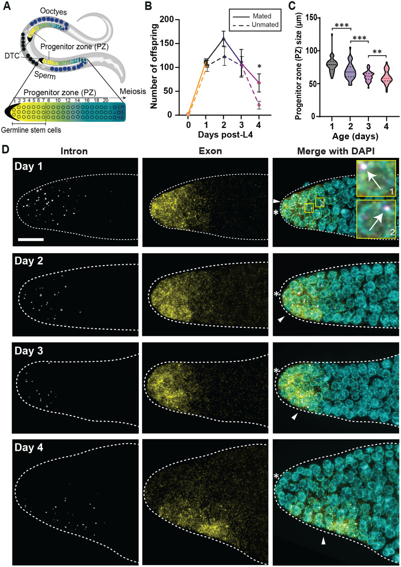
Age-induced decline in germline function begins early in adulthood whereas the germ cells age minimally. (A) Schematic of adult C. elegans hermaphrodite with U-shaped gonads with a distal tip cell (DTC) at each distal end (black crescents). Along the proximal end, oocytes (dark blue) reside that meet with sperm to form eggs (black). Germline stem cells (GSCs; yellow) occupy the first 6–8 germ cell rows of the germline. The GSCs progress through the progenitor zone, begin differentiation (green) to oocytes by entering meiosis (blue). (B) Egg laying rate for mated (solid line) and unmated (dashed line) N2 hermaphrodites from mid-L4 to Day 4. (C) The size of the progenitor zone (PZ), which measures the distance between the distal end of the gonad and the first cells in meiotic prophase (crescents). n=45 gonads. (B,C) All error bars in this study are the standard error of the mean (SEM) unless stated otherwise. For all t-tests (one sample or two sample) in this study, *P<0.05, **P<0.01, ***P<0.001, and ****P<0.0001. ‘n.s.’: not significant by t-test. (D) sygl-1 smFISH with the wild type, N2, at different aging stages. Day 1: the young adult stage (24h post mid-L4), Day 2: 48h post L4, Day 3: 72h post L4, and Day 4: 96h post L4. The distal gonads are shown in Z-projection. Left: the intron probes reveal sygl-1 nascent transcripts at the active transcription sites (ATS). Middle: the exon probes show both the nascent transcripts (bright dots) and cytoplasmic mRNAs (small, dim dots). Right: DAPI marks DNA in the nucleus. The arrow indicates ATS, which is seen both in the intron and exon channel. White asterisk: distal end of the gonad. Arrowhead: the position of the DTC/niche nucleus. Scale bar: 10 µm, the area in the numbered yellow boxes are shown 10× zoomed in on the right.

