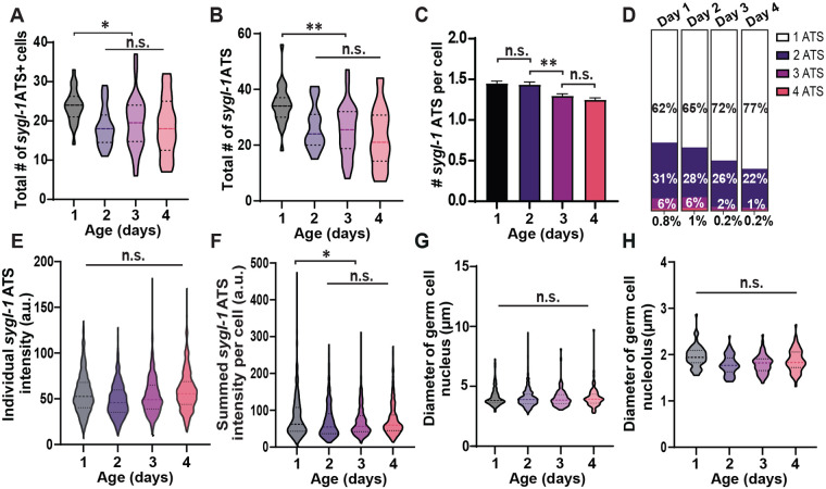
Fig. 2.
Aging reduces sygl-1 transcriptional activation both at the tissue and cellular levels. (A) The total number of sygl-1 ATS-containing cells in the gonad for each aging stage. For violin plots in this study, the middle line shows the median; top and bottom dashed lines are the third and first quartiles. (B) The total number of sygl-1 ATS in the gonad. (C) The average number of sygl-1 ATS in each germ cell for all ages. (D) The sygl-1 ATS-containing cells are grouped by the number of ATS they contain and plotted as the percentages for each age. (A–D) n=27, 25, 26, and 24 gonads for Day 1, 2, 3, and 4 respectively (E) The individual sygl-1 ATS intensities are compared between all ages. n=621, 460, 508, and 423 nuclei for Day 1, 2, 3, and 4 respectively. (F) The signal intensities of all sygl-1 ATS in each germ cell are pooled to calculate the summed sygl-1 ATS intensity, which estimates the overall sygl-1 transcriptional activity in each cell. n=931, 661, 660, and 550 ATS for Day 1, 2, 3, and 4 respectively. (E,F) a.u.: arbitrary units. (G) The diameter of the germ cells located at the distal gonad (0–40 μm from the distal end of the gonad) was measured for each aging stage. n=3043, 2645, 2703, and 2662 nuclei for Day 1, 2, 3, and 4 respectively. (H) The diameter of the nucleolus in each germ cell located at the distal gonad (0–40 μm from the distal end) was recorded for all ages. n=65 nucleoli for all ages.

