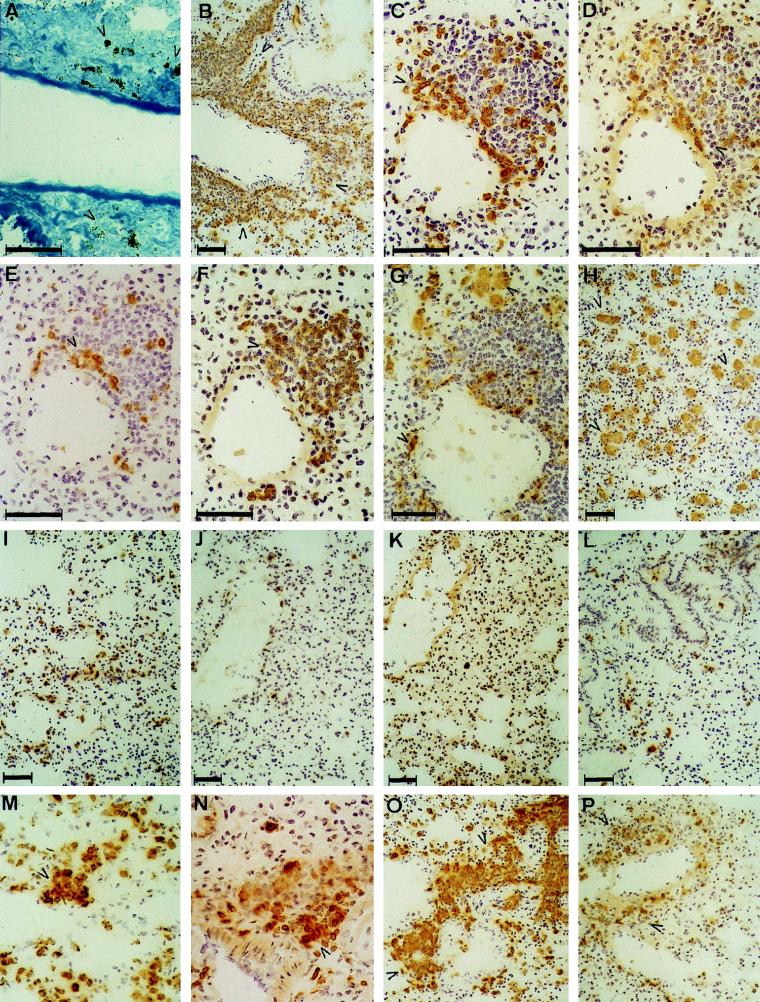
FIG. 5.
Histopathology of the lungs in invasive murine candidiasis at day 3 in GKO (A to H) and C57BL/6 mice (I to L). Inflammatory lesions in GKO mice at day 2 (M and N) and day 14 (O and P) are also shown. Perivascular infiltrates appeared only in GKO mice and contained MHC II+ (TIB120+) mononuclear cells (B), CD3+ T cells (C), mostly CD4+ (D) and fewer CD8+ (E) cells and B220+ B cells (F), which were also CD19+ (not shown). Mφ FA/11+ (G) were not a dominant component of the infiltrates, but were present in two different phenotypes in the lungs: elongated, perivascular cells and engorged, diffusely distributed alveolar Mφ (H). Perivascular infiltrates contained only debris of C. albicans, as shown by Gomori’s staining (A). Histopathology of the lungs in IL-4 KO and C57BL/6 mice was markedly different from that of GKO mice; a similar inflammatory reaction did not develop in these strains even at later stages of infection. Lung sections obtained at day 3 from C57BL/6 mice were stained with TIB120 (I), FA/11 (J), B220 (K), and anti-CD3 (L) MAbs and showed only minimal inflammatory responses. At day 2, perivascular MHC II+ cell aggregates (M) appeared in GKO mice, most of which were CD3+ T cells (N), and by day 14, the extension of perivascular infiltrates was reduced compared with that of day 3, containing MHC II+ cells (O), which were partly CD3+ T cells (P). Bars, 50 μm.

