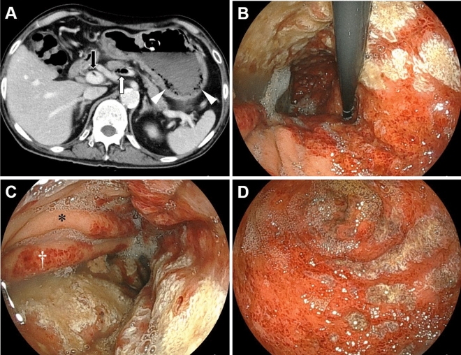
Figure 2.
CT and endoscopic images of a 66-year-old male, currently undergoing chemotherapy for esophageal cancer, with a gastrostomy tube placement. The patient was asymptomatic, and during an endoscopic evaluation before esophageal cancer surgery, gastric mucosal injury was noted. On the same day, CT scans (A) revealed the presence of gas (white arrow) and thrombosis (black arrow) in the splenic to portal veins. Additionally, a gas-filled appearance was observed within the gastric wall (arrowheads). Esophagogastroduodenoscopy (B–D; B, the gastric fornix and upper body; C, gastric body; D, gastric antrum) showed diffuse reddish erosions with white exudates extending from the fornix to the antrum. The boundary between the injured and non-injured mucosa was partially distinct (B). The anterior wall of the gastric body was intact (C, asterisk). Mucosal injury was predominant on the folds (C, dagger). CT computed tomography.

