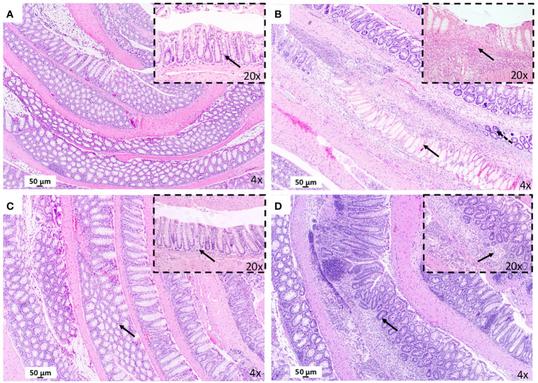
Figure 2.
Treatment with maqui extract improve histopathological evaluation of colonic tissue from animal model of Crohn’s disease-like colitis after Hematoxylin and Eosin staining (H&E). A representative image from entire “Swiss rolled” colon evaluation from the distal to proximal end at 40x magnification (objective 4) (n= 6 mice/group, see Supplementary Figure S2 ). (A) Control Group: Normal histology colon tissue. Physiology immune cells in lamina propria (black arrow in 10x). (B) CD Group: Presence of necrosis (black arrow in 40x). and goblet cells depletion (dashed arrow). Transmural infiltrate inflammatory (black arrow in 20x). (C) post-Treatment group: Preservation of the colonic mucosa structure (black arrow in 40x). Normal cells infiltration (black arrow in 20x). (D) pre-Treatment group: Lesser extent of atrophy (black arrow in 4x) and mucosal crypts loss (black arrow in 20x).

