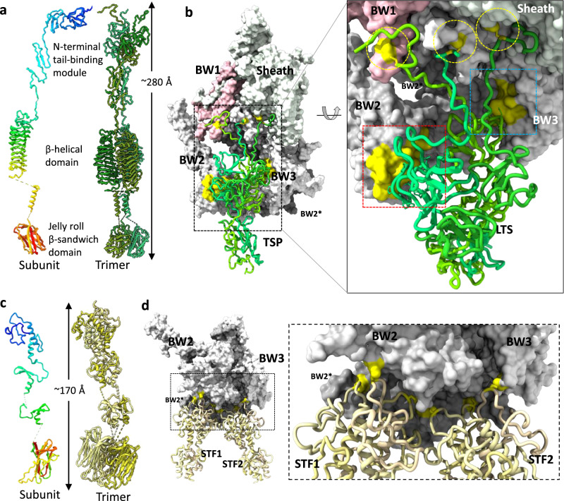
Fig. 5. Structural organization of Mialno receptor binding proteins.
a Structure of tail spike (TSP, gp124) subunit and trimer. The N- to C-terminus of the subunit is colored blue-to-red. b Interactions of TSP and baseplate components. TSP and baseplate components are shown in the green ribbon model and surface representation, respectively. The TSP interactions with baseplate are highlighted by yellow surface and circles. The cysteine residues involved in disulfide bonds are colored and circled yellow. Detailed protein–protein interactions at red and blue-squared interfaces are shown in Supplementary Fig. 9. c Structure of short tail fibers (STF, gp31) subunit and trimer. The N- to C-terminal of subunit is colored blue-to-red. d Interaction of STF and baseplate components. STF and baseplate components are shown by tan ribbon model and surface representation, respectively. The cysteine residues involved in disulfide bonds are colored yellow.

