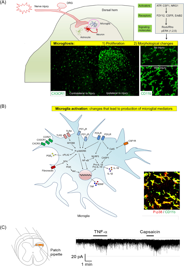
Figure 10:
Microglia in the development of neuropathic pain. (A) Nerve injury-induced microgliosis (proliferation and morphological changes). (B) Nerve injury-induced microglia activation, as indicated by upregulation and activation of ATP receptors, chemokine receptors, and TLRs (TLR4 and TLR2) [171, 184–188] and phosphorylation of MAP kinases p38 and ERK (1/2, 5) [169, 189–192]. As a result of microglia activation, pro-inflammatory mediators, such as TNF, IL-1β, IL-18, and PGE2, as well as growth factor (BDNF), are produced and secreted from microglia, inducing central sensitization in the spinal cord and enhancing pain states [151, 193–195]. A and B are reproduced from Chen et al. [42] with CCC permission. (C) Patch clamp recording in spinal cord slice reveals rapid increase in excitatory post-synaptic currents in spinal cord pain circuit following TNF treatment. The same neuron also responded to TRPV1 agonist capsaicin. Reproduced from Park et al. [196] with permission (author’s own right).

