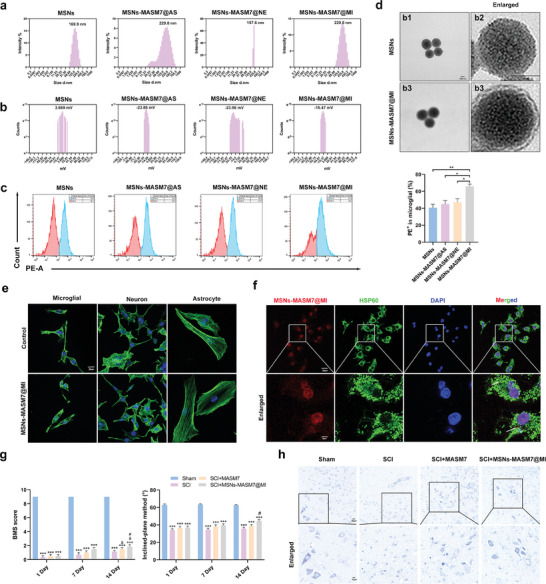
Figure 7.

Nanomaterials carrying MASM7 promoted neuroprotection and functional recovery after SCI. a) The diameter distribution of MSNs, MSNs‐MASM7@AS, MSNs‐MASM7@NE, or MSNs‐MASM7@MI. b) The zeta potential distribution of MSNs, MSNs‐MASM7@AS, MSNs‐MASM7@NE, or MSNs‐MASM7@MI. c) Flow cytometric analysis of nanomaterials (coated by microglia membranes, neuron, and astrocyte membranes) uptake by primary microglial. d) TEM images of the MSNs and MSNs‐MASM7@MI (Scale bar = 50 nm). e) Fluorescence images of cellular morphology (F‐actin) in primary microglial, neuron, and astrocyte cells after 24 h (Scale bar = 20 µm). f) Fluorescence images of MSNs‐MASM7@MI and mitochondria (HSP60) in primary microglial after 24 h (Scale bar = 20 µm). g) Effects of different treatments on neurological function scores at 1, 7, and 14 d after SCI. h) Nissl staining in the perilesional tissues 7 d after SCI (Scale bar = 20 µm).
