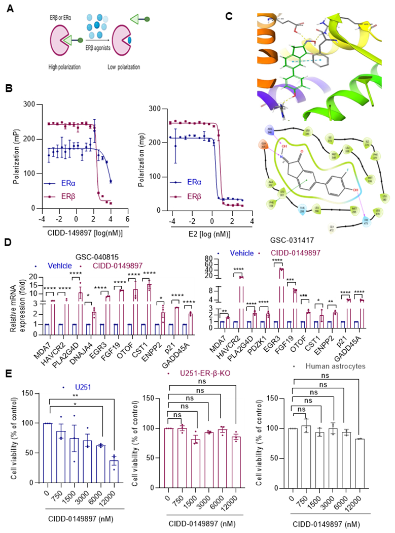
Figure 2.

Target specificity of CIDD-0149897. A, Schematic of polar screen ER binding assay. B, Specificity of E2 (left panel) and CIDD-0149897 (right panel) to ERα and ERβ determined using polar screen ER competitive binding assays. Fluorescence polarization is measured and shown as millipolarization (mP) (n=2) C, Induced fit docking pose of CIDD-0149897 in the ligand binding site of PDB structure 1X7B. (Top panel) 3D rendering of docking pose indicating hydrogen bonding (yellow) and pi-stacking (cyan) interactions. (Bottom panel) 2D representation of ligand interactions. D, Effect of CIDD-0149897 treatment (10 μM, 72h) on ERβ targeted genes was measured using RT-qPCR analysis in GSC-040815, GSC-031417cells (n=3). E, Effect of CIDD-0149897 on normal human astrocytes as well as on WT and ERβ-KO U251 GBM cells was measured using the MTT cell viability assay (n=3). Data are presented as mean ± SEM * p<0.05, ** p<0.01, *** p<0.001, ****p<0.0001. In D, p-values were calculated using two-way ANOVA. In E, p-values were calculated using one-way ANOVA.
