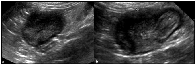
Figure 4.
Transverse ultrasound image of (a) the ascending colon and (b) ileocolic junction in a 3-year-old female cat with feline infectious peritonitis (case 10). (a) The ascending colon is thickened with lost wall layering (between the calipers: 8.5 mm). (b) An ileocecocolic junction-centered colonic wall thickening with loss of wall layering (between the calipers: 8 mm). The ileum is also seen (asterisk)

