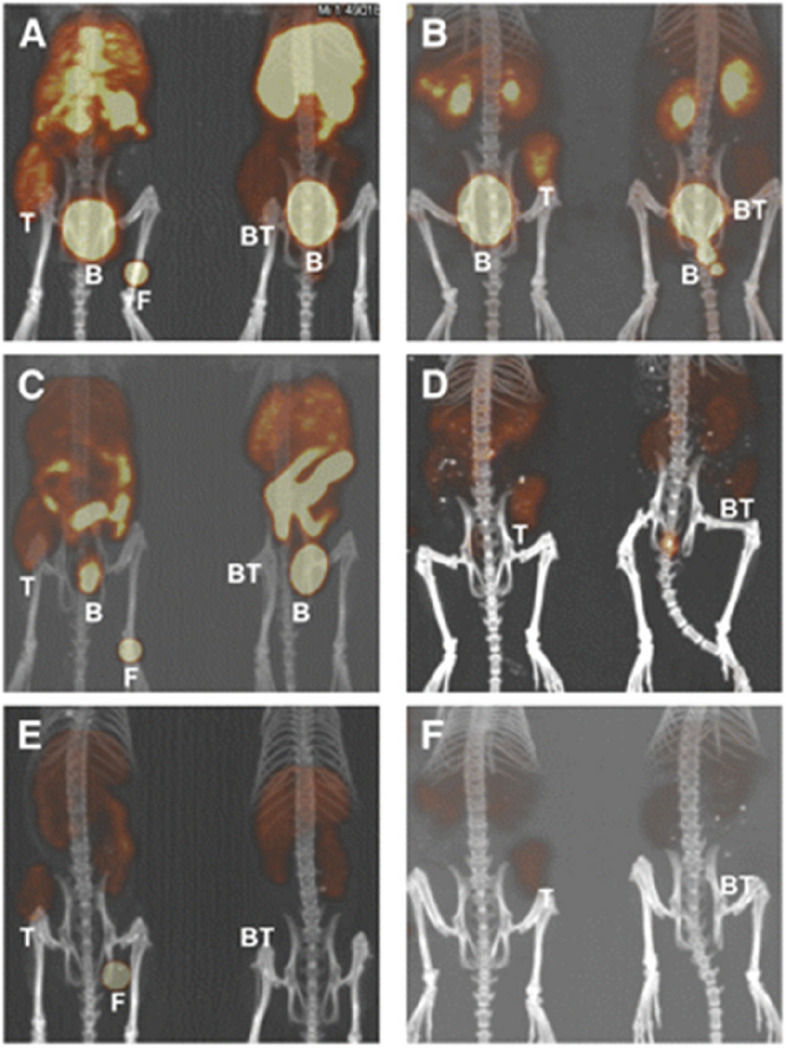
Fig. 2.

Coronal views of maximum-intensity projections of small-animal PET images with coregistered CT image of mice bearing PC-3 xenografts in rear flank at 1 (A and B), 4 (C and D), and 24 h (E and F). Mice were injected intravenously with 64Cu-SarAr-SA-Aoc bombesin(7–14) (A, C, and E; tumors on the left flank) and 64Cu-SarAr-SA-Aoc-GSG bombesin(7–14) (B, D, and F; tumors on the right flank). Mice on the left of each frame were not injected with a blocking agent, whereas mice on right received 100 µg of Tyr4-bombesin as an inhibitor. Fiducial markers (F) are also shown in some images. B = bladder; BT = blocked tumors; T = nonblocked tumors. This study was originally published in JNM. Lears, K.A., et al., In vitro and in vivo evaluation of 64Cu-labeled SarAr-bombesin analogs in gastrin-releasing peptide receptor-expressing prostate cancer. J Nucl Med, 2011. 52(3): p. 470–7. © SNMMI
