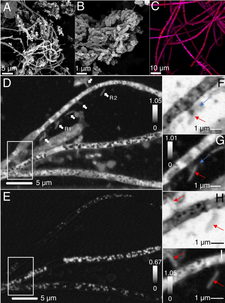
Fig. 2.

Microscopic characterization of MS4 biofilms. A–B Scanning electron microscopy of filamentous bacteria and associated cells, small cells are pointed by arrows (C) confocal fluorescence microscopy of cells treated with SYTOX (blue) for nucleic acid and F-64 (red) for membrane. Scanning transmission x-ray microscopy: (D) carbon map of filaments and associated cells (white arrows). E Corresponding distribution map of S0 evidencing spherical elemental sulfur granules within the compartments of the filaments. The top, middle, and bottom filament widths are 1.23 ± 0.5 µm, 1.01 ± 0.2 µm, and 1.33 ± 0.3 µm, respectively. F An ultra-small cell ~ 480 nm long, ~ 270 nm wide, (blue arrow) in contact with an apparently episymbiotic cell 1.86 ± 0.1 µm long, ~ 360 nm wide (red arrow), imaged at 280 eV (region R1, D) and corresponding (G) carbon map. H Two apparently episymbiotic cells (red arrows) connected to filaments, imaged at 280 eV (region R2, D) and corresponding (I) carbon map. The intensity scales correspond to optical density
