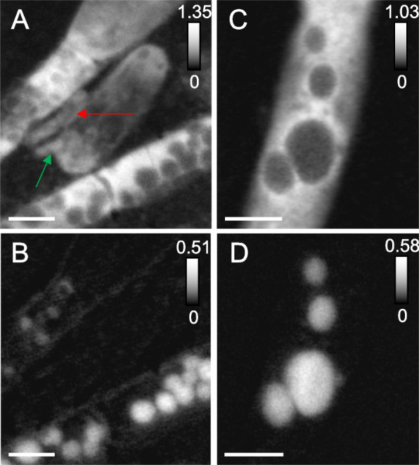
Fig. 3.

Scanning transmission X-ray microscopy of MS4 and MS11 biofilms. A Protein map and corresponding (B) distribution map of S0 in MS4 biofilms (in white boxed area of Fig. 2). Cells that are 908 ± 32 nm long, 370 ± 30 nm wide (red arrow), 687 ± 34 nm long, 244 ± 33 nm wide (green arrow), seen in close contact with filaments. C Protein map and corresponding (D) distribution map of S0 in MS11 biofilms, showing the presence of sulfur granules (up to 1.08 ± 0.12 µm in diameter) in a small area of a long filament. Sulfur L2,3 edge spectra of the granules can be found in Fig. S2. The intensity scale corresponds to the optical density. Scale bars: 1 µm
